



Blog
Achilles Tendinopathy (Tendonitis): Expert Treatment Guide

Written by: James Gill BOst GOsC 6039. Registered Osteopath, Founder & Clinic Director

Medically Reviewed: Lauren Egginton MChiro GCC 06282, Registered Chiropractor. Specialist in Pregnancy, Paediatric Chiropractic & Functional Rehabilitation | London.
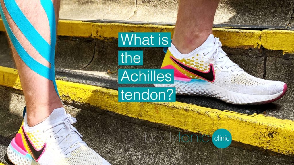
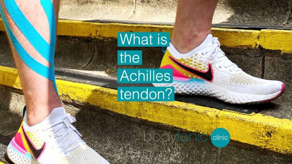
What is the Achilles tendon?
A tendon is a band, made of connective tissue, connecting a muscle to the bone. A tendon can be “stretched”, “strained” or even torn, but not “sprained” (you use this terminology for a ligament, as opposed to “strained” which is specific to a muscle or tendon).
The Achilles tendon (or calcaneal tendon) is located in the posterior (back) aspect of the lower leg, attaching the gastrocnemius, soleus and plantaris muscles to the calcaneus bone. This group of muscles serves in the plantar flexion of the foot (bringing the front foot towards the ground) and flexion of the knee. The Achilles tendon is the thickest in the human body (Doral et al., 2010).
Andrew O’Neill: “The Achilles is designed to be a spring, not a cable. When patients lose that ‘springiness’ due to thickening or inflammation, we focus on restoring the elastic capacity of the tendon, not just stretching it out.”
Tendons have different functions depending on their location: they can be “positioning” or “energy storing”, the latter being the case of the Achilles tendon for example (Thorpe et al., 2013). Energy storing tendons store and recover energy during a specific motion, such as during the human stride. An imbalance in this storing/recovering system can lead to the tendon’s irritation, or worse, a tendon tear which can be partial or full.
Causes of Achilles Tendinopathy
A tendonitis (or tendinopathy) can happen due to overloading, repetitive strain, or trauma. The most common context leading to Achilles issues is running. Factors include:
- Return to activity after inactivity.
- Postural imbalance affecting the posterior chain.
- Poor footwear or restricted foot mobility.
- Biomechanical issues often linked to Plantar Fasciitis.
We separate the lower limb into two chains: posterior (gluteals, hamstrings, calves) and anterior (hip flexors, quads, tibialis). A healthy balance is key to preserving the integrity of the muscles and their tendons.
Ensure your equipment is fully functional
A tendonitis (or tendinopathy) can have various causes and can happen in different contexts. An irritation of the Achilles tendon can happen due to overloading, repetitive strain, or even a trauma, which can lead to a full rupture of the tendon. The most common context leading to Achilles tendonitis is usually running, and multiple factors can explain it: return to activity after a period of inactivity, postural imbalance leading to a stress of the posterior chain muscles of the leg (glutei, hamstrings and calves), poor shoewear support, or restricted foot mobility.
We commonly separate the muscle group of the lower limb in two chains: posterior and anterior. The posterior group is composed of the gluteals, hamstrings and calves, whereas the anterior is composed of the hip flexors, quadriceps and tibialis muscles. A healthy balance in the loading and activation of these chains will be key to preserving the integrity of the muscles and their tendons.
Clinical Insight from James Gill, BOst | Founder & Clinic Director: “At our Canada Water and Stratford clinics, we often find that the Achilles is overworking because the gluteal muscles aren’t providing enough propulsion. By addressing the entire ‘Kinetic Chain,’ we ensure the tendon isn’t just healed, but protected from future overload.”
Achilles Tendonitis Infographic


What are the typical symptoms of Achilles tendonitis?
How can you diagnose an Achilles tendonitis?
While the signs and symptoms described by the patient will help in the diagnosis of an Achilles tendonitis and lead the physical examination, the only way to get a precise and clear medical diagnosis is through imaging.
The best devices to visualise soft tissues like tendons will be MRI and ultrasound machines. They will give a very detailed image of the integrity of the Achilles tendon, and in the case of an ultrasound, will even show a real-time image of this structure in motion, which will help understanding the mechanism leading to the symptoms described by the patient. An X-Ray won’t visualise the soft tissues, such as muscles, ligaments, tendons, but will highlight other structures more dense such as bones. Therefore, an X-Ray will help rule out other conditions that could create similar symptoms, but won’t be effective diagnosing a tendonitis.
How long does an Achilles tendonitis last?
Untreated and with repetitive activity designed to stress the tendon, such as running or jumping (especially if wearing an inappropriate shoewear that is poorly supporting the foot, and/or on a concrete surface), the symptoms of the Achilles tendonitis may take a few weeks or even months before feeling better. With the appropriate self care and even treatment options (as listed below), the symptoms may only take 2 to 3 weeks before dissipating. However, it is always recommended to consult a specialist to avoid a repetition of these inflammations.
With appropriate Specialist MSK Treatment, symptoms may dissipate in 2–3 weeks. However, chronic cases require targeted intervention to avoid rupture.
With appropriate Specialist MSK Treatment, symptoms may dissipate in 2-3 weeks. However, chronic cases require targeted intervention to avoid the risk of a full rupture.
What are the treatment options for Achilles tendonitis?
There are many options to treat Achilles tendonitis: you can manage it yourself or you can see a practitioner.
- Ice vs. Heat: Where the use of heat is helpful to treat muscle tightness, here ice would be more beneficial as it will be an effective tool to calm down the inflammatory process on the tendon. Because of the potential skin reactions when in direct contact with heat or ice, it is always recommended to use a tissue or a thin cloth wrapped around the source of cold before applying it on the sore area. An application on the painful location every 3 to 4 hours, for not more than 15-20 mins at a time should be enough.
- Rest is also recommended in the first 2-3 days following the initial presentation of the symptoms, to reduce the pain level and minimise the stress applied to the tendon, but cannot be considered as a mid- or long-term solution as it won’t treat the reason leading to this irritation.
- If after trying self management for a few weeks the symptoms persist, an examination and assessment by a specialist would be recommended. The first step for further medical referral would be to consult a GP, who could refer the patient to a physiotherapist or to an orthopaedic specialist. In some tendonitis cases, steroid injections may be recommended, providing a short term pain relief, but these can’t be performed in the case of Achilles tendonitis. The reason for it is because it may weaken the tendon and put it at risk of further damage.
- Specialist Clinical Care: Consulting an Osteopath, Chiropractor or MSK physiotherapist in the treatment of an Achilles tendinopathy will provide the benefits of a comprehensive postural assessment, which will give you a clear and reasonable understanding of the mechanism leading to Achilles tendonitis. Treatment will aim to work globally on the lower extremity, not only the patient’s foot and ankle, but also the knee, hip, pelvis and further up in order to handle the cause of the symptoms and not only their presentation.
Storz Medical Shockwave Therapy
If rest and physical therapy fail to resolve your pain after several weeks, Shockwave Therapy is the most effective non-surgical step for chronic Achilles Tendonitis. We utilise the versatile Radial Shockwave (ESWT) to treat the broad tendon structure and accelerate healing, and the precision of Focused Shockwave (FSWT) to target deep insertion pain or any associated calcific deposits, ensuring the most complete and rapid return to activity.
What are the best exercises for Achilles tendonitis?
An appropriate loading of the tendon, associated with a comprehensive and reasonable plan of return to activity discussed with the patient will be key to enhance the healing process and avoid any risks of relapse.
1. Isometric Contraction
When loading an irritated tendon, it’s preferable to do it with what is called an “isometric” contraction: the muscle is contracted, but not lengthened or shortened. The exercise can be done standing up, with the possibility to find your balance by placing one hand on an elevated support, such as a desk. Whilst a weight (potentially a dumbbell) is held in one hand, you stand on one leg on the front of your foot, tip-toes, and hold the position for 15-20 seconds. Repeat the exercise 5 times, with a 40 seconds break in between repetitions. This exercise can be done every day as a part of a muscle activation program, and specifically before any physical activity such as running or jumping.
2. Ankle and Foot Mobility
Another type of exercise will be designed to stabilise the ankle and foot mobility. This will ensure the tendons and ligaments in the area are loaded appropriately, with a good absorption of the ground forces by the joints of the foot and ankle on each step of a run, or when landing after a jump.
The 4 key movements that will be reinforced with this exercise are the plantar flexion, the dorsiflexion, the inversion and eversion of the foot. With an elastic band pulling in the opposite direction of the movement done, either with a hand hold of the elastic or wrapped to a stable object, you will need to do the motion against the resistance, and then gradually release your traction for a return to a neutral position. Effectively, this exercise will allow a concentric (shortening) and eccentric (lengthening) contraction of the muscle targeted.
3. Return to Activity
Finally, to plan carefully a return to activity, it is recommended to do lower impact type of runs, preferably on an even soft surface or grass if possible. Running for a smaller distance and at a slower pace in the first 10 days of the program. This will allow the tendon to heal, be strengthened, whilst still active. These exercises are also vital for Ankle Sprain Rehab.
Clinical Insight from James Gill: “A common mistake is returning to running on flat pavement too soon. We often suggest our Stratford patients use the softer trails in Queen Elizabeth Olympic Park for their initial return-to-run phase to reduce the peak impact on the tendon.”
Can Achilles tendonitis be prevented?
Because all of the exercises quoted previously are designed to avoid further repetitions of the Achilles tendonitis, they can both be seen as rehab but also prehab tools: a good balance, stability of the ankles and enough mobility in the feet will keep the risk of tendonitis at a low level. This is why consulting a specialist for routine check-ups and maintenance Sports Massage will put the body in the best conditions to function optimally and avoid unnecessary stress!
Insertional Achilles Pain
Insertional Achilles pain is a specific type of Achilles tendinopathy where the irritation occurs at the point where the tendon attaches to the heel bone (calcaneus). This is slightly different from mid-portion Achilles tendinopathy, which affects the tendon a few centimetres above the heel. Insertional pain often requires a slightly modified approach to treatment and exercise.
Symptoms:
- Localised pain at the back of the heel, often aggravated by wearing shoes that rub against the area.
- Morning stiffness and pain when taking the first steps of the day.
- Pain with activities that load the tendon, such as running, jumping, or going up stairs.
Treatment options:
Management of insertional Achilles pain involves a combination of conservative and specialist therapies:
- Osteopathy, Chiropractic, and Physiotherapy: Hands-on assessment and treatment help address not only the tendon but also foot, ankle, knee, and hip mechanics to reduce stress on the Achilles.
- Shockwave Therapy: High-energy sound waves can be applied to promote healing, stimulate collagen production, and reduce pain at the tendon insertion.
- Modified Exercise Programmes: Eccentric and isometric loading exercises are tailored to avoid excessive compression at the heel. Often, exercises are performed on a flat surface rather than on tiptoes to reduce stress at the insertion point.
- Footwear & Orthotics: Using shoes with a heel lift or cushioned support can reduce tension on the tendon during activity.
Activity Modification: Reducing high-impact activities and gradually reintroducing load is crucial for healing.
Clinical Insight from Mr Andrew O’Neill: “Insertional cases require patience. Because the tendon is compressed against the bone, we often use ‘Flat-Ground Loading’ rather than dropping the heel off a step. This prevents further irritation while still building the necessary strength for recovery.”
Key point: Insertional Achilles tendinopathy tends to be slower to heal than mid-portion Achilles tendonitis due to the tendon’s attachment and compressive forces at the heel. Patience, correct loading, and specialist guidance are essential for full recovery.
References
Doral, M.N., Bozkurt, M., Turhan, E., Dönmez, G., Demirel, M., Kaya, D., Ateşok, K., Atay, Ö.A. and Maffulli, N., 2010. Achilles tendon rupture: physiotherapy and endoscopy-assisted surgical treatment of a common sports injury. Open access journal of sports medicine, 1, p.233.
Thorpe, C.T., Birch, H.L., Clegg, P.D. and Screen, H.R., 2013. The role of the non‐collagenous matrix in tendon function. International journal of experimental pathology, 94(4), pp.248-259.
Osteopathy, MSK Physiotherapy & Chiropractic Prices
New Clients: Save 10% on your 1st Massage, Beauty, or Wellness treatment* Excludes Medical & Clinical services.
Axa / Bupa Insurance Accepted*